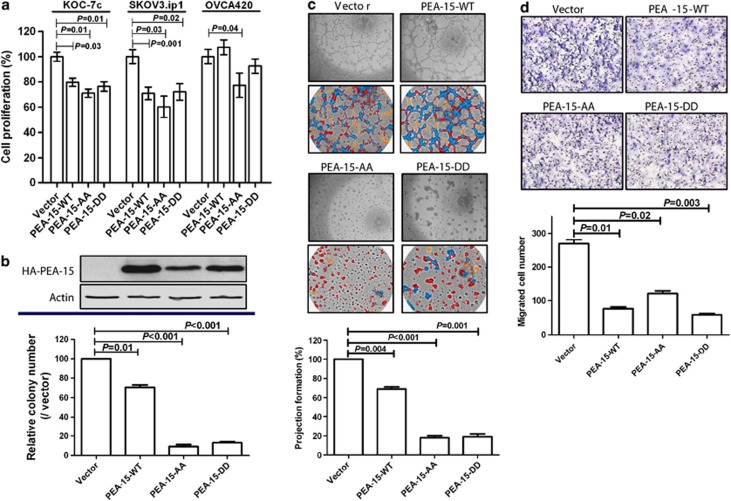
Figure 2.
PEA-15-AA and PEA-15-DD inhibited cell proliferation and anchorage-independent growth. (a) Cells were transiently transfected with vector, PEA-15-WT, PEA-15-AA or PEA-15-DD for 72 h, and then cells (5 × 10⁴) were plated into a six-well plate. After 72 h, cell numbers were counted by trypan blue staining. (b) In vitro tumorigenicity (soft agar) assay. Stably transfected SKOV3.ip1 cells (2 × 10³) were seeded into soft agar and incubated for 3 weeks. Colonies were stained with 200 ml of iodo-nitrotetrazoliumchloride (1 mg/ml) for overnight, and the number of colonies over 80 mm in diameter was counted using the GelCount colony counting system according to the manufacturer's instructions. (c) Three-dimensional Matrigel culture assay. Cells (2 × 10⁴) were added onto the μ-Slide plate (ibidi GmbH) according to the manufacturer's instructions. Blue indicates nodal structure; yellow, well-developed tube formation; red, poorly developed tube formation. (d) Migration assay. Cells (1 × 10⁵) were added into the upper well and 10% FBS was added into the lower well as an attractants. After a 6-h incubation, migrated cells were stained and counted. Columns, mean; bars, standard deviation. Statistical significance was evaluated by paired t-test using GraphPad Prism software. Data shown are representative of three experiments with similar results.

