Figure 4.
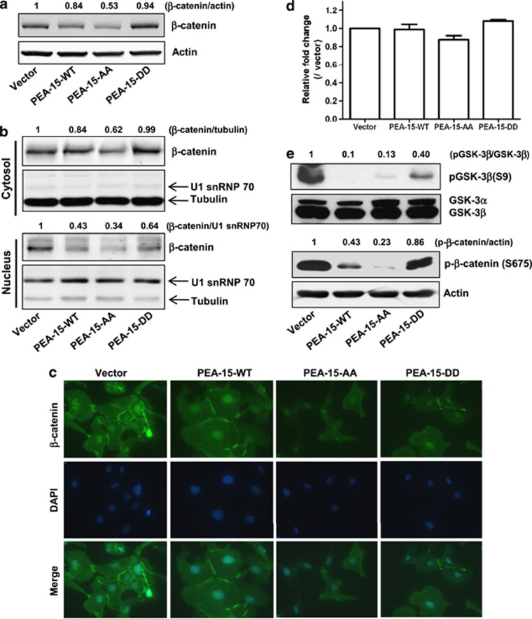
PEA-15-AA inhibited β-catenin expression and translocalization. (a) Total β-catenin expression level in stably transfected SKOV3.ip1 cells. Fifteen micrograms of lysate was loaded. The expression levels of β-catenin for each cell line were evaluated using LI-COR software. (b) PEA-15-AA inhibited translocalization of β-catenin into the nucleus. Cytosolic and nuclear fractions were prepared as described in Materials and methods. For loading control, tubulin (for cytosolic fraction) and U1 snRNP 70 (for nuclear fraction) were used. (c) Immunofluorescence staining. Stably transfected SKOV3.ip1 cells were grown on the four-well slide chamber for overnight, and immunofluorescence staining was performed as described in Material and methods. (d) Quantitative RT–PCR analysis of β-catenin mRNA level in stably transfected SKOV3.ip1 cells. (e) Western blotting. Effect of different forms of PEA-15 on GSK-3β phosphorylation at Ser9 and β-catenin phosphorylation at Ser675.

