Figure 4.
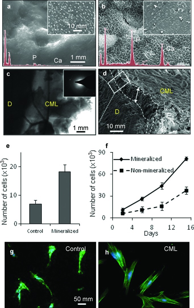
Structural and functional characteristics of the cementomimetic layer formed on the root of human tooth by ADP5. (a and b) SEM images of the demineralized (a) and (b) ADP5-formed cementomimetic layer revealing uniform nanocrystals with a Ca/P ratio of 1.67 obtained from EDX () spectra (insets). (c) TEM images and the electron diffraction pattern of the newly formed cementomimetic mineral layer in cross-section showing HAp crystallites. (d) SEM image of mechanically separated cementomimetic mineral layer displaying uniform thickness of crystallized HAp. (e) Attachment of hPDL cells on control and cementomimetic mineral layer. (f) Proliferation of the hPDL cells on control, uncoated, root stock compared to ADP-induced cementomimetic mineral layer. (g and h) Fluorescent microscopy image showing F-actin. Cell attachment without formation of organized actin network on control surface (g) is compared to those on ADP-induced cementomimetic mineral layer that reveals a well-organized actin cytoskeleton and lamellapodia (h). EDX, energy dispersive X-ray; HAp, hydroxyapatite; hPDL, human periodontal ligament; SEM, scanning electron microscopy; TEM, transmission electron microscopy.

