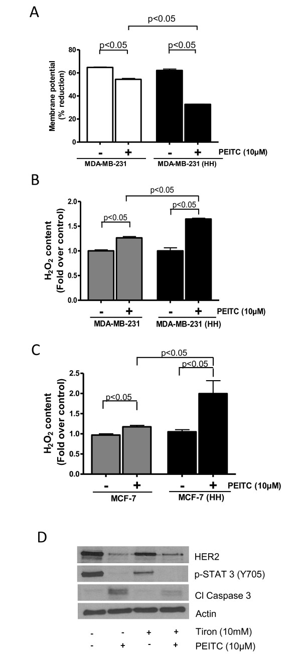
Figure 7.
(PEITC) causes ROS generation and mitochondrial depolarization. (A) MDA-MB-231 and MDA-MB-231 (high HER2 (HH)) cells were treated with PEITC for 24 h and then were labeled with tetramethylrhodamine and analyzed by flow cytometer (n = 3). Hydrogen peroxide content was measured in control or 10 μM PEITC-treated (B) MDA-MB-231 and MDA-MB-231 (HH) cells and (C) MCF-7 and MCF-7 (HH) cells. (D) MDA-MB-231 cells were treated with 10 mM Tiron followed by treatment with 10 μM PEITC for 24 h. The cell lysate was analyzed for HER2 expression. Statistically different when compared with control (P < 0.05).

