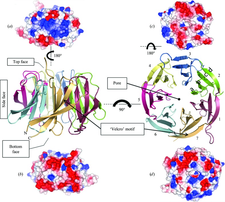
Figure 2.
Views of the hRack1 crystal structure. Central panel: cartoon representations of hRack1 viewed from the side (left) and from the top (right). The locations of the protein pore and the ‘velcro’ motif are indicated. Each β-sheet or blade is numbered sequentially from the N-terminus of the protein and their β-strands are labelled a, b, c, and d starting from the inside of the propeller near the pore. The electrostatic surface of hRack1 is shown from four different angles. Side surfaces, bottom and top views are displayed in (a) (blades 1–3), (b) (blades 5–7), (c) and (d), respectively.

