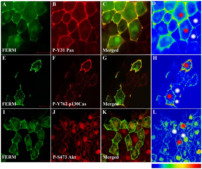
Figure 6. The FERM domain activates endogenous FAK leading to increased phosphorylation of FAK/Src targets.
(A–D) Embryos injected with HA-FERM mRNA in two blastomeres, at the animal pole, at the four cell stage were processed for immunofluorescence using anti-HA (green) and anti-P-Y31 paxillin (red) antibodies. C is the merged image and D is an intensity color coded image. FERM expressing cells display elevated levels of phosphorylated paxillin (red stars) when compared with un-injected neighbouring cells (white stars). (E–H) Same as (A–D) but comparing phosphorylation levels of p130Cas on tyrosine 762 between FERM expressing and control cells. FERM expressing cells show elevated levels of phosphorylated p130Cas (red stars), when compared with un-injected cells (white stars). (I–L) Same as (A–D) but comparing levels of phosphorylated Akt on serine 473 between FERM expressing and control cells. Levels of phosphorylated Akt are comparable in FERM expressing cells to those of control neighboring cells. Scale bars: 20 µm.

