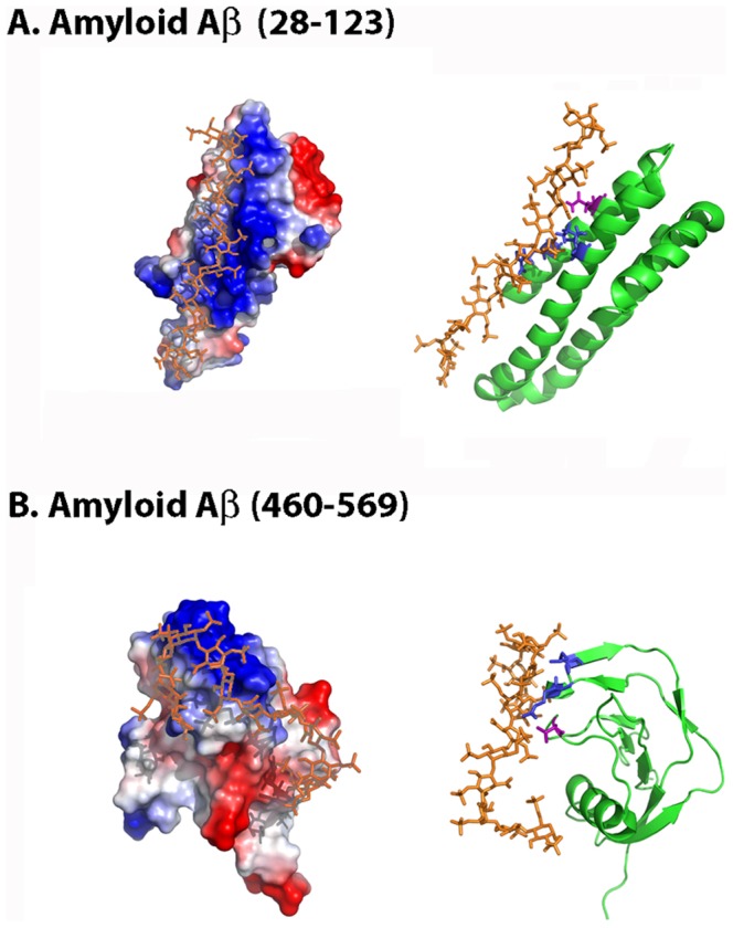
Figure 5. Molecular docking simulation of Aβ28–123 and Aβ460–569 heparin-binding sites.

The figure displays the protein electrostatic potential (left) and the protein cartoon highlighting in red the CPC clip motif (right) of (A) Aβ28–123 and (B) Aβ460–569. CPC residues are colored in blue (cationic) and magenta (polar). Heparin dodecasaccharide ligand used in docking simulations is colored in orange. PDB codes: 1MWR (Aβ28–123), 1TKN (Aβ460–569) and 1HPN (heparin ligand).
