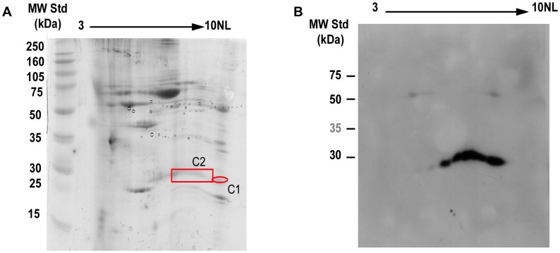
Figure 3. Identification of putative natural self antigen.
A) Bidimensional electrophoresis gel stained with colloidal Coomassie Brilliant Blue. Sixty five µg of purified carotid atherosclerotic plaque proteins were loaded on strip pH 3-10NL, 7 cm. The 2nd dimension was carried out using 12.5% SDS-PAGE. B) 2D electrophoresis Western Blotting. Similarly, 70 µg of proteins were loaded on strip pH 3-10NL, 7 cm. The 2nd dimension was carried out using 12.5% SDS-PAGE. After transfer, proteins were probed with Fab7816-FLAG (10 µg/mL). Protein spots of interest (red box) were excised from the gel, digested with trypsin and analysed by MALDI-ToF mass spectrometry. In both spots human TAGLN was identified with almost complete sequence coverage.

