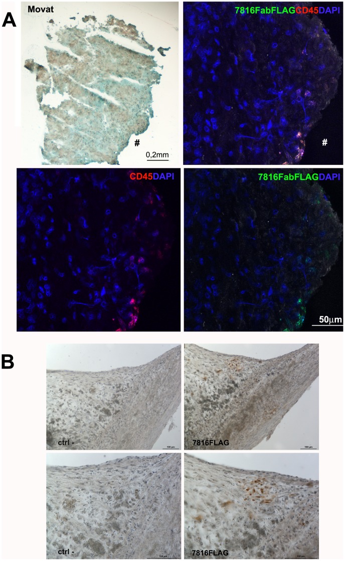
Figure 4. Immunofluorescence on human coronary plaqes and Immunohistochemistry on human carotid sections with Fab7816-FLAG.
a) Representative section stained by Movat’s pentchrome (left panel) showed the morphology of a portion of the coronary plaque tissue (plaque ID-A) displaying slightly damaged media rich in smooth muscle cells. Confocal microscopy (right panels) showed the presence of CD45+ cells (red) labelled by Fab7816-FLAG revealed by MAb M2 anti-FLAG-FITC (green) in the coronary plaque tissue region indicated by symbol (#). DAPI stained the nuclei (blue). b) Immunoperoxidase on carotid plaque samples demonstrated the presence of several cells reacting with Fab7816-FLAG in an area close to the lumen (left panels) as revealed by MAb M2 anti-FLAG-HRP developed with DAB (brown). The signal is absent in a serial section where the Fab7816-FLAG is omitted (ctrl-, right panels). Magnified images in the bottom panels evidence the spindle shape of Fab7816-FLAG+ cells and their localization in between a clusters of altered cells, possibly foam cell. Haematoxylin (blue) stains nuclei. Scale bars indicate the magnification.

