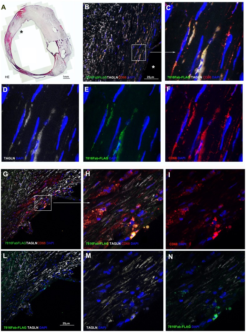
Figure 5. Confocal microscopy on human atherosclerotic carotid sections.
Multiple staining of two carotid plaques are displayed to characterize the Fab7816-FLAG+ cells type. First plaque A-F panels, 2nd plaque G-I to N. The reconstruction of a section from 1st plaque stained with haematoxylin and eosin (HE) obtained by multiple images grabbing tool of Lucia-G software is in A; asterisk indicates the lumen in correspondence of Fab7816-FLAG+ cells, on the shoulder of the atheromasic lesion demonstrated in B by confocal lmicroscopy. Confocal microscopy images in B,C and G,H demonstrated by Fab7816-FLAG (green), goat-anti-human TAGLN (white), mouse-anti-human CD68 (red) the presence of triple positive cells in the intima, closely to the lumen. Single or double staining are shown in D-F and I-N. Squares and arrows indicated the enlarged areas. DAPI stains the nuclei (blue). Scale bars indicate the magnification.

