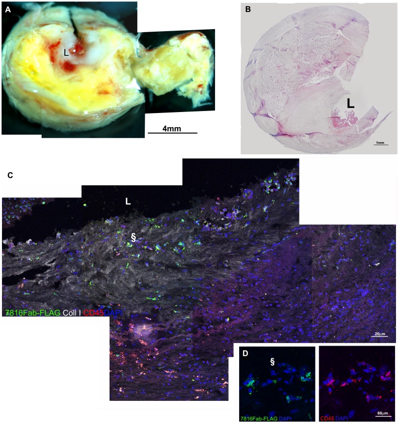
Figure 6. Confocal microscopy on human atherosclerotic carotid sections.
The macroscopic aspect of a carotid plaque is reconstructed in A by stereomicroscope and displays a lipidic core and of luminal thrombus. Features are confirmed in B by histology (haematoxylin and eosin). Confocal microscopy in C shows a reconstruction of part of the area close to the lumen (L) in a section stained with Fab7816-FLAG (green), mouse-anti-human Collagen type I (white) and mouse anti-human CD45 (red), revealed by opportune secondary antibodies. In D is an enlargement of the area indicated by § symbol in C with several Fab7816-FLAG+/CD45+ cells in the neointima. indicate a regions magnified. Scale bars indicate the magnification.

