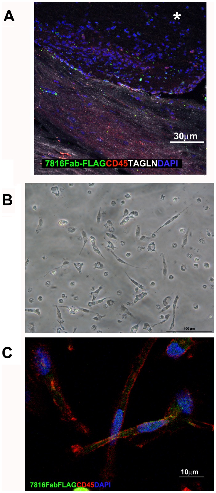
Figure 7. Fibrocytes morphology and immunoreactivity with 7816Fab FLAG.
A) Confocal microscopy staining showing presence of 7816Fab FLAG +/CD45+/TAGLN+ cells in human carotid plaque lesion. B,C) Confocal microscopy image of non-confluent CD14+ fibrocytes grown on glass for 4 days in the absence of serum. Spindle shaped cells are stained with 7816Fab FLAG +/CD45+(green and red, respectively). DAPI stains the nuclei (blue). Scale bars indicate the magnification.

