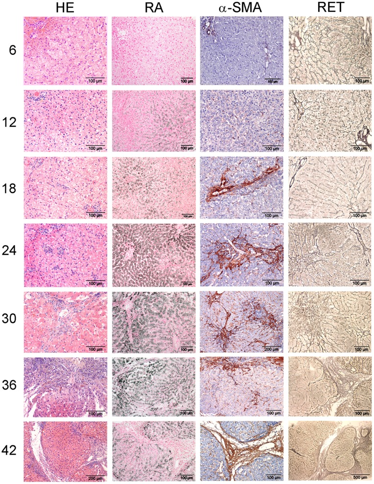
Figure 1. Histological description.
COMMD1-deficient dog livers were stained with H&E and RA to assess inflammation and copper accumulation, respectively, and stained for α-SMA and reticulin (collagen type III) to assess fibrosis. Representative pictures of a COMMD1-deficient dog over a period of 42 months are shown. Numbers indicate age in months.

