
Figure 1.
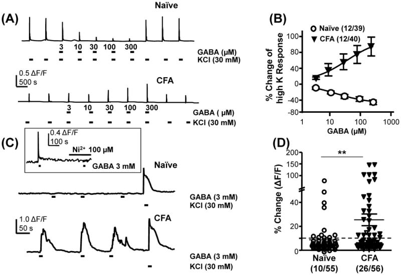
Inflammation-induced increase in the excitatory actions of GABA in cutaneous DRG neurons. A) High K (30 mM KCl) was used to evoke Ca2+ transients in neurons from naïve and inflamed (CFA) rats. Co-application of GABA with high K results in a concentration dependent suppression of the Ca2+ transient in a subpopulation of neurons from naïve rats, but an increase in the transient in a subpopulation of neurons from inflamed rats. B) Pooled data from neurons in which GABA increased (CFA) or decreased (Naïve) high K evoked transients. C) Direct application of GABA to cutaneous neurons from naïve rats resulted in little change in the intracellular Ca2+ concentration in the majority of neurons tested. However, in close to half of the neurons from inflamed rats, direct application of GABA resulted in a transient increase in intracellular Ca2+. High K was used to confirm neurons could respond to a depolarizing stimulus. Inset, GABA evoked Ca2+ transients were completely blocked by 100 μM Ni2+. D) Pooled data tested with direct activation of GABA. The proportion of cutaneous neurons from inflamed rats directly activated by GABA was significantly greater than that from naive rats. ** p < 0.01, χ2 test.