
Figure 7.
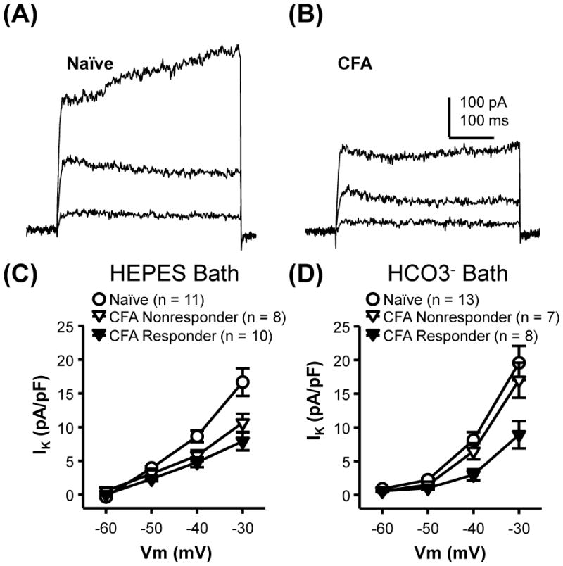
Inflammation-induced decrease in outward current in cutaneous neurons. Typical non- or weakly-inactivating outward current evoked with voltage steps to −50, −40 and −30 mV from −60 mV in cutaneous neurons from naïve (A) and inflamed (B) rats. The currents showed in B, were from a neuron that responded to GABA with a Ca2+ transient. Note the absence of low threshold inward current in either in A or B which was typical of cutaneous neurons studied from naïve and inflamed rats. C) Pooled data from naïve rats and responders and nonresponders from inflamed rats collected in HEPES buffered bath solution. D) Pooled data from the same 3 groups of neurons collected in HCO3- buffered bath solution. For data plotted in both C and D, two-way mixed design ANOVA revealed a significant influence of voltage (p < 0.01) and group (p < 0.01), as well as a significant interaction between the two. Post-hoc analysis indicated that current density in responders from inflamed rats evoked at −40 and −30 mV was significantly smaller that than in neurons from naïve rats.