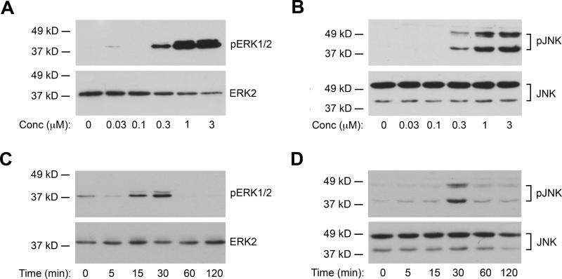
Fig. 2. Chalcone-24 stimulates ERK1/2 and JNK activity in A549 cells.
(A) and (B) A549 cells were incubated for 30 minutes with the indicated concentrations of chalcone-24. Whole cell lysates were analyzed by immunoblot for (A) phosphorylated, active ERK1/2 (top panel, 20 μg protein) and total ERK2 (bottom panel, 10 μg protein) or (B) phosphorylated, active JNK1 and JNK2 (top panel, 40 μg protein) and total JNK (bottom panel, 10 μg protein). (C) and (D) A549 cells were incubated for the indicated times with 0.3 μM chalcone-24. Whole cell lysates were analyzed by immunoblot for (C) phosphorylated, active ERK1/2 (top panel 30 μg protein) and total ERK1/2 (bottom panel, 10 μg protein) or (D) phosphorylated, active JNK1 and JNK2 (top panel, 40 μg protein) and total JNK (bottom panel, 10 μg protein). The data shown are representative of at least two independent experiments.

