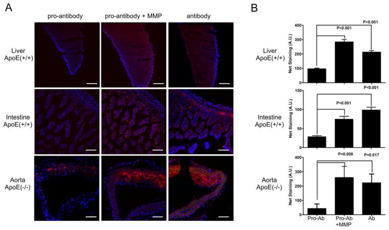
Figure 3.
Analysis of anti-VCAM-1 antibody and pro-antibody binding to explanted tissue sections using fluorescence microscopy. (A) Tissue sections from ApoE(+/+) (liver, intestine) and ApoE(−/−) (aortas) mice were stained with Alexa546-conjugated anti-VCAM-1 antibody or pro-antibody (100 nM), before and after treatment with MMP-1. (Red - Alexa546;– Blue). Original magnification ×20, Scale bars, 100 μm.. (B) Explanted tissues were stained with Alexa546-conjugated anti-VCAM-1 antibody or pro-antibody (50 nM). Bars represent average and standard error of the integrated fluorescence signals from 4–6 fields for each tissue. Significance was confirmed using both Student’s t-test (shown), and one-way ANOVA analysis with Bonferroni’s correction. However, the staining signal difference in the aorta between the pro-antibody and antibody samples did not reach statistical significance as determined using Bonferroni’s multiple comparison test.

