Figure 6.
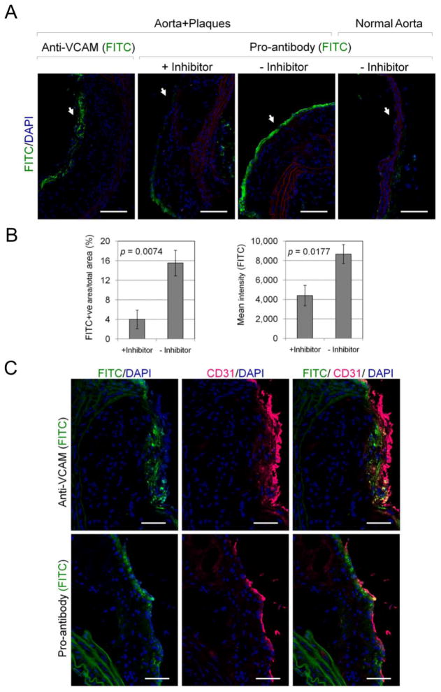
Pro-antibody binding to aorta explants in the presence and absence of MMP inhibitor. (A) Cross-section of aortas comparing the bound FITC-labeled monoclonal VCAM-1 antibody and pro-antibody within luminal endothelial surface of plaque tissue (arrow; green). Original magnification ×20. Scale bars, 100 μm. (B) Quantitative analysis of pro-antibody binding to plaques in the presence and absence of MMP inhibitor expressed as FITC-positive area / total selected area (Mean % ± SE; from n=3 mice/group), and mean fluorescence intensity (Mean ± SE; from n=3 mice/group). (C) Localization of endothelial cell marker (anti-CD31, magenta) and anti-VCAM-1 antibody or pro-antibody-FITC (green) in aorta sections. Original magnification ×40. Scale bars, 50 μm. Significance was confirmed using Student’s t-test (shown) and a Mann-Whitney test.

