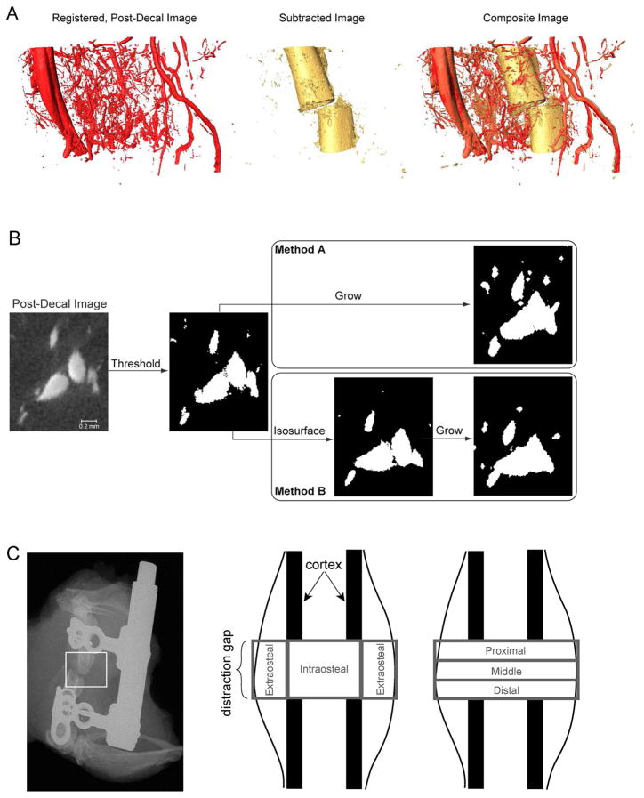
Figure 1. Methods for using μCT to co-localize and to analyze vascular and mineralized tissues within the distraction regenerate and the surrounding musculature.
(A) Shown at the left is a rendering of the vasculature (POD 14) after image registration was carried out on the post-decalcification image in order to remove distortion of the tissues that occurs when the mineral is removed. Subtraction of this registered image from the pre-decalcification image produces an image of the mineralized tissue alone (shown in the middle). Examination of the composite image of bone and vasculature (shown on the right), obtained from overlaying the subtracted image on the pre-decalcification image, allows analysis of the spatial relationship between vascular and osseous tissues. Even though plain-film, radiographic assessments will show optimal alignment before the device is removed, some slippage and misalignment can occur when the bone is embedded in agarose, as seen with the reconstructed images. (B) As represented on a small region of a 2-D cross-section of one image, the process of image subtraction involved first binarizing the registered post-decal image in order to segment the vasculature and then growing the vessels by a small amount prior to subtracting the image from the pre-decal image. The two growth methods that were evaluated are illustrated for a growth value of 3. For the second method (method B), the isosurface representation of the vessels is shown in the intermediate step to illustrate how the isosurface is smoother than the original vessel boundaries determined purely by thresholding; growth is applied to the isosurface rather than the original boundaries. (C) Two types of regional analysis were carried out on the portion of the regenerate, inclusive of surrounding musculature, located between the two osteotomy cuts. In the first, the regenerate was partitioned into intraosteal and extraosteal volumes of interest (VOIs), and in the second the partitioning was into proximal, middle, and distal VOIs. The union of each of these two sets of VOIs was dubbed the “total VOI”.

