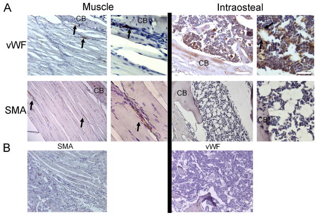
Figure 4. Identification of Vascular Structures in Un-Operated Control Tissues.
(A) Selected micrographs (100x and 400x) of immunohistological analysis of smooth muscle actin (SMA; associated with arteriogenesis) and von Willibrand factor (vWF; associated with angiogenesis) in muscle and bone tissue from the femoral mid-diaphyseal region are shown. Arrows indicate immune positive structures, and ‘CB’ indicates cortical bone. White scale bars are of length 200 and 50 microns, respectively, for the low- and high-magnification images. (B) Micrographs (100x) of control sections incubated with species-appropriate, non-immune serum and secondary antibody conjugates

