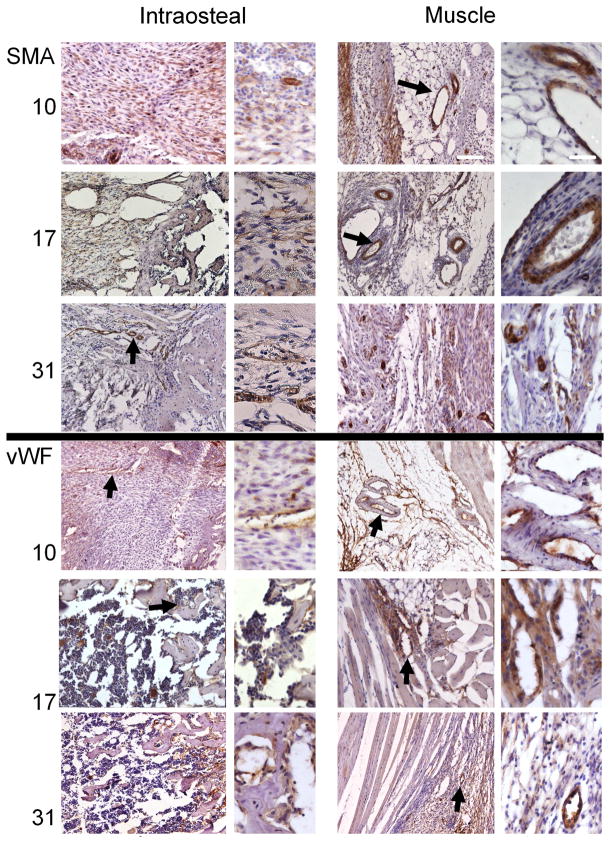
Figure 5. Immunohistological assessments of vessel formation during distraction osteogenesis.
Selected micrographs (100x and 400x) of immunohistological analysis of smooth muscle actin (SMA; associated with arteriogenesis) and von Willibrand factor (vWF; associated with angiogenesis) were taken from muscular areas surrounding the distraction gap and from the intraosteal region, as defined by the periosteal margin, at days 10, 17, and 31. Time-points are listed in the left margin. Bold arrows indicate immune-positive structures that are shown in greater detail in the adjacent higher-magnification images. The micrographs showing intraosteal regions are always oriented so that the long axis of the bone is displayed horizontally, with the proximal direction at the left. The micrographs showing muscle tissues are oriented so that the diaphysis, which is vertically oriented, is to the right of the field of view. White scale bars are of length 200 and 50 microns, respectively, for the low- and high-magnification images.

