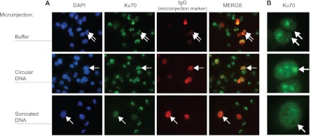
Fig. 5.
DNA ends are sufficient to induce exit of Ku from the nucleolus in vivo. A, MDA-MB-231 cells were microinjected with rabbit immunoglobulin (IgG) as an injection marker, alone (top row) or in combination with circular (second row) or sonicated (third row) plasmid DNA and processed for immunofluorescence following a triton extraction and paraformaldehyde fixation protocol (T/PFA). Cells were labeled with an anti-rabbit IgG antibody to identify microinjected cells (red channel) and with an anti-mouse IgG antibody to detect endogenous Ku70 (green channel). Nuclei were stained with DAPI (blue channel). B, Cropped views of microinjected cells from (A) showing highly magnified nuclei. Arrows indicate representative microinjected cells.

