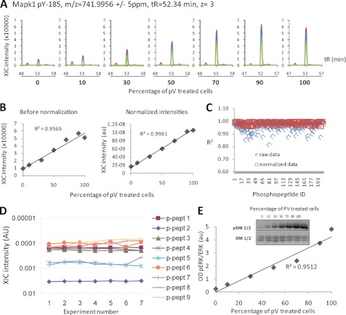
Fig. 1.
Accuracy and precision of phosphopeptide quantification by label-free LC-MS/MS. Extracts of pV-treated cells were mixed with extracts of untreated cells at the proportions shown in A, followed by enrichment of phosphopeptides and quantification by LC-MS. A, XICs of a phosphopeptide matching MAPK Tyr(P)-185 in samples containing increasing amounts of pV-treated cells. XICs of the first, second, and third isotopes are shown in blue, red, and green, respectively. B, plot of data, as returned by Pescal, demonstrating the linearity of quantification before and after normalization. C, the linearity of the quantitative data was confirmed by the analysis of 200 additional phosphopeptides induced upon pV treatment. After normalization, R2 > 0.95 for each of the analyzed phosphopeptides was achieved. D, plot of XIC intensities of nine phosphopeptides in seven replicates demonstrating the precision (reproducibility) of the analysis. E, aliquots of lysates from the experiment shown in A–C were analyzed by immunoblotting using an antibody against MAPK Tyr(P)-185 (ERK1/2). The plot shows that phosphosite quantification by immunoblotting is not as linear as quantification by LC-MS (compare E with B). OD, optical density.

