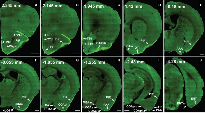
Figure 10.
The ventral lateral MOB projects to olfactory cortical structures from the AON to the ENT (2.345 to –4.28 mm from bregma). Axons ramify in AONd, AONm, AONpv (A), TTd, TTv (B,C), PIR (A–I), OTl (D), AAA (E), NLOT (F), COAa (F–H), COApl (G–I), BA (G), MEAav (H), PAA (H,I), TR (I), and ENTl (J) without any spatial topography. The DP, OTm, COApm, and ENTm do not receive inputs from the ventral lateral MOBmi (B,D,I,J). The PIR-OT junction contains dense labeling (C), where the fibers appear to wrap around layer II pyramidal neurons in OTl (Figures 12H,I). Axon numbers appear to decrease as they travel medially (B–J) and caudally (A–J). Differences between projection patterns from the ventral lateral and dorsal medial MOBmi were not detected (see Figure 11). Case number SW101213-01A. Scale bar, 500 μm. Abbreviations: AONd, dorsal anterior olfactory nucleus; AONm, medial AON; AONpv, posterior ventral AON; TTd, dorsal taenia tecta; TTv, ventral TT; PIR, piriform area; OTl, lateral olfactory tubercle; OTm, medial OT; AAA, anterior amygdalar area; NLOT, nucleus of lateral olfactory tract; COAa, anterior cortical amygdalar area; COApm, posterior medial COA; BA, bed nucleus of accessory olfactory tract; MEAav, anterior ventral medial amygdalar nucleus; PAA, piriform-amygdalar area; TR, postpiriform transition area; ENTl, lateral entorhinal area; ENTm, medial ENT.

