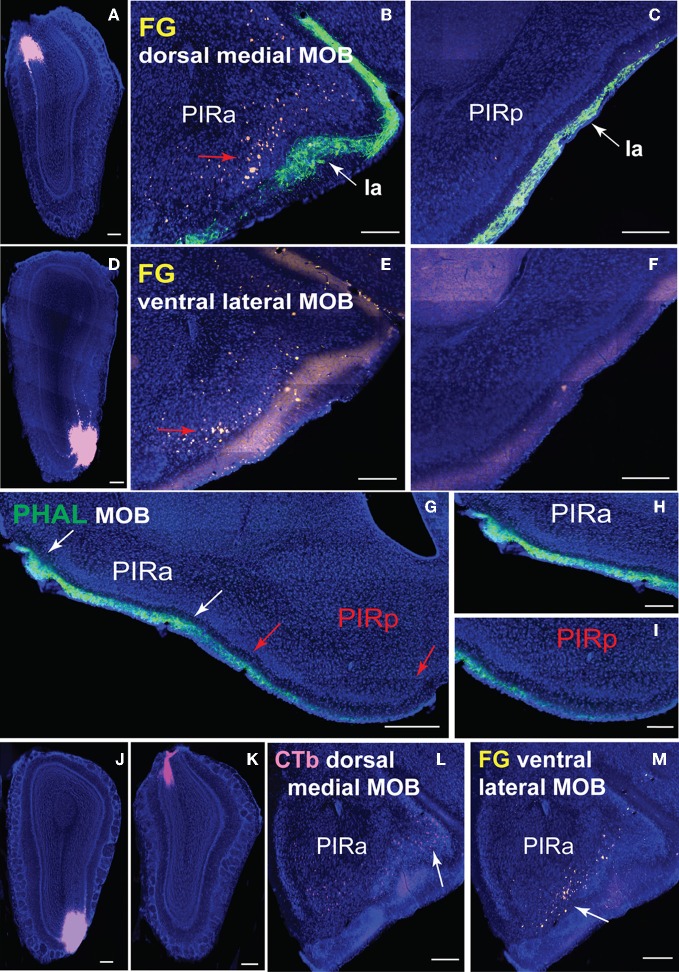
Figure 9.
Connectional differences between PIRa and PIRp. FG injections in dorsal medial (A) and ventral lateral (D) MOBmi back-label neurons in PIRa (B,E), but not in PIRp (C,F). Layer Ia of PIR, labeled with PHAL (see Figure 6A2 for PHAL injection), appears to get thinner in width from PIRa to PIRp (B,C,E–I). Input to PIRa and PIRp is also different as MOB projects more densely to PIRa compared to PIRp (G–I). PIRa between white arrows (G) is magnified in (H), while PIRp in between red arrows (G) is magnified in (I; see Figures 1A,B for PHAL injection). Topographic arrangement of neurons in PIRa (red arrows in B,E) are more clearly observed when FG and CTb are double injected in dorsal versus ventral MOBmi, respectively (J,K). Dorsal MOBmi projecting CTb neurons occupy more dorsal regions of PIRa (L), while ventral projecting neurons are in more ventral parts of PIRa (M). Scale bar, 200 μm; 500 μm (G). Case numbers SW101213-01A (A–C), SW101215-03A (D–F), SW101215-02A (G–I), SW110607-03A (J–M).

