Figure 1.
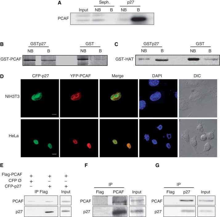
Interaction of p27 with PCAF. (A) HCT116 cell extracts were loaded on a p27 or a Sepharose 4B column (Seph.). Proteins not bound (NB) or bound (B) to the columns were analyzed by WB using an anti-PCAF antibody. (B) Purified GST–PCAF was loaded on a GST–p27 or on a GST column. Fractions not bound (NB) or bound (B) to the columns were visualized by gel electrophoresis and Coomassie blue staining. (C) Purified GST–HAT was loaded on a GST–p27 or a GST column. Fractions not bound (NB) or bound (B) to the columns were visualized by gel electrophoresis and coomassie blue staining. (D) NIH3T3 and HeLa cells were cotransfected with CFP–p27 and YFP–PCAF, stained with DAPI and subsequently visualized by confocal microscopy. Bar: 5 μm (E) HEK293T cells were transfected with Flag–PCAF or/and CFP–p27. At 24 h post-transfection, cell extracts were immunoprecipitated with anti-Flag. Cells transfected with an empty CFP vector (Ø) and Flag–PCAF were used as a control. The immunoprecipitates were then analyzed by WB using antibodies against PCAF or p27. (F) HEK293T cell extracts were subjected to IP with anti-PCAF or anti-Flag (as a control), then, the immunoprecipitates were analyzed by WB using antibodies against PCAF or p27. (G) HEK293T cell extracts were subjected to IP with anti-p27 or anti-Flag (as a control), then, the immunoprecipitates were analyzed by WB using antibodies against PCAF or p27.

