Figure 5.
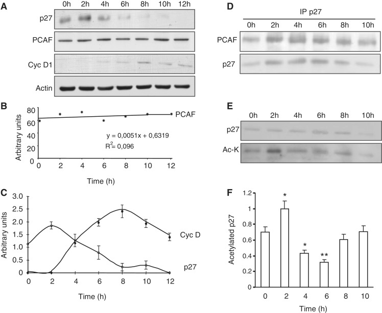
The p27 protein is acetylated at early G1. (A) HeLa cells were made quiescent by serum starvation for 72 h and then re-plated in the presence of 10% FCS. Total cell extracts were prepared at the indicated times after cell-cycle resumption. The levels of PCAF and p27 were examined by WB with anti-PCAF and anti-p27. WB with anti-cyclin D1 was performed to monitor cell-cycle progression and WB with anti-actin was performed as a loading control. (B) PCAF levels from three different experiments described in (A) were quantified and expressed as a regression line. (C) cyclin D1 and p27 levels from three different experiments described in (A) were quantified and expressed as the mean value ± SD. (D) HeLa cell extracts from the indicated times were subjected to IP with anti-p27 and the levels of PCAF and p27 in the immunoprecipitates were determined by WB. (E) Extracts from HeLa cells at different times after cell-cycle resumption were subjected to IP with anti-p27. Then, the levels of p27 and those of acetylated p27 in the immunoprecipitates were assessed by WB with anti- p27 or with anti-acetyl-lysine (Ac-K), respectively. (F) The acetylation levels of p27 from three different experiments described in (E) were quantified and expressed as the mean value ± SD. Statistical analyses were performed using Student's two-tailed paired t-test. *P < 0.05, **P < 0.01.

