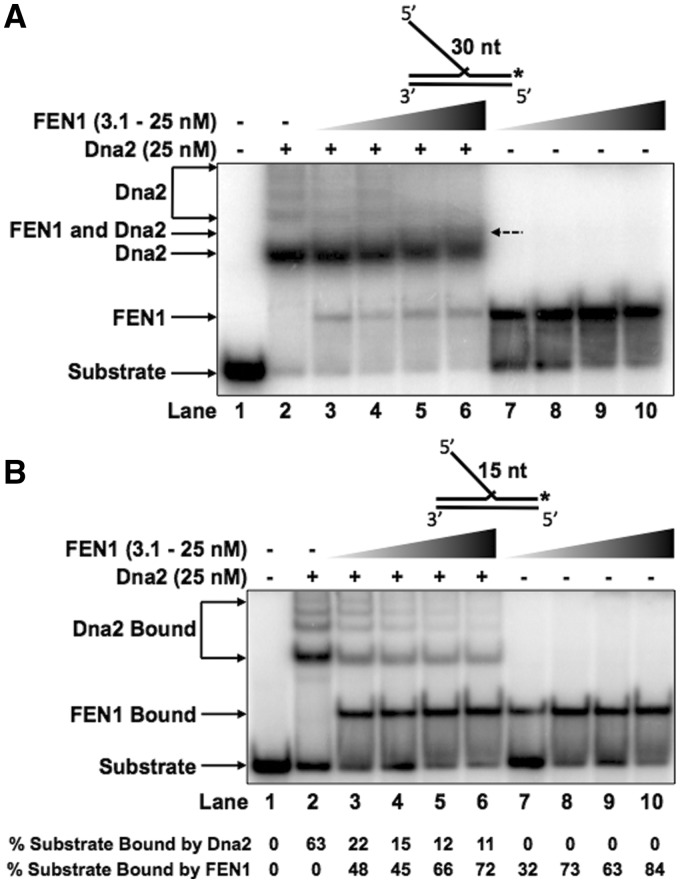
Figure 4.
FEN1 preferentially displaces Dna2 from intermediate length double-flap structures. Dna2 (25 nM) was pre-incubated with the experimental substrate prior to the addition of increasing concentrations of FEN1 (3.1, 6.25, 12.5 or 25 nM in lanes 3–6). Lane 1 shows the substrate alone, lane 2 shows Dna2 bound without FEN1 and lanes 7–10 show FEN1 alone bound to the substrate at the same concentrations as in 3–6. (A) shows Dna2 and FEN1 binding competition for the 30 nt double-flap substrate (U2:T3:D2.30). (B) shows Dna2 and FEN1 binding competition for the 15 nt double-flap substrate (U2:T3:D2.15). In (A) and (B) the position of the substrate alone, Dna2–substrate complex and FEN1–substrate complex are indicated to the left of the figure. In (A), the FEN1–Dna2–substrate complex is indicated to the left of the figure and to the right of lane 6 with a dashed arrow. In (B), the quantitation of the percent substrate bound by hDna2 or hFEN1 is shown below the figure.

