Fig. 1.
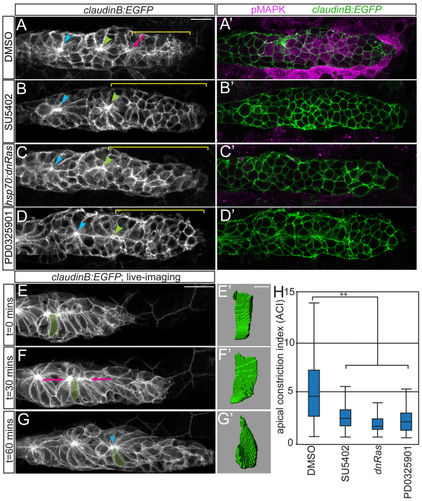
Fgfr-Ras-MAPK signaling in the pLLp is required for rosette formation. (A-D) Confocal projections showing rosette formation in the DMSO control (A) and with Fgfr (B), Ras (C) and MAPKK (D) inhibition in claudinB:EGFP zebrafish embryos at 30 hpf. Arrowheads indicate centers of the trailing rosettes. Pink arrow indicates nascent rosette. Brackets indicate rosette-free region. (A′-D′) pMAPK immunolabeling in single planes from projections in A-D. Fgfr and MAPKK inhibition with 100 μM SU5402 and 7 μM PD0325901, respectively, was from 28-30 hpf. Ras inhibition via hsp70:dn-Ras induction was at 28 hpf; embryos were fixed 2 hours following heat shock. (E-G) Stills from time-lapse movie of pLLp in a wild-type claudinB:EGFP embryo (supplementary material Movie 1). (F) Cells align apical ends along the midline (pink arrows). (G) Center of the nascent rosette (arrowhead). (E′-G′) Three-dimensional reconstructions of the highlighted cell in E-G. (H) ACIs for embryos treated with DMSO, SU5402, PD0325901 or following induction of hsp70:dn-Ras. n=180 cells from six embryos per condition. **P<0.0001, Wilcoxon test. Error bars indicate s.e.m. Scale bars: 20 μm in A-G; 4 μm in E′-G′.

