Fig. 2.
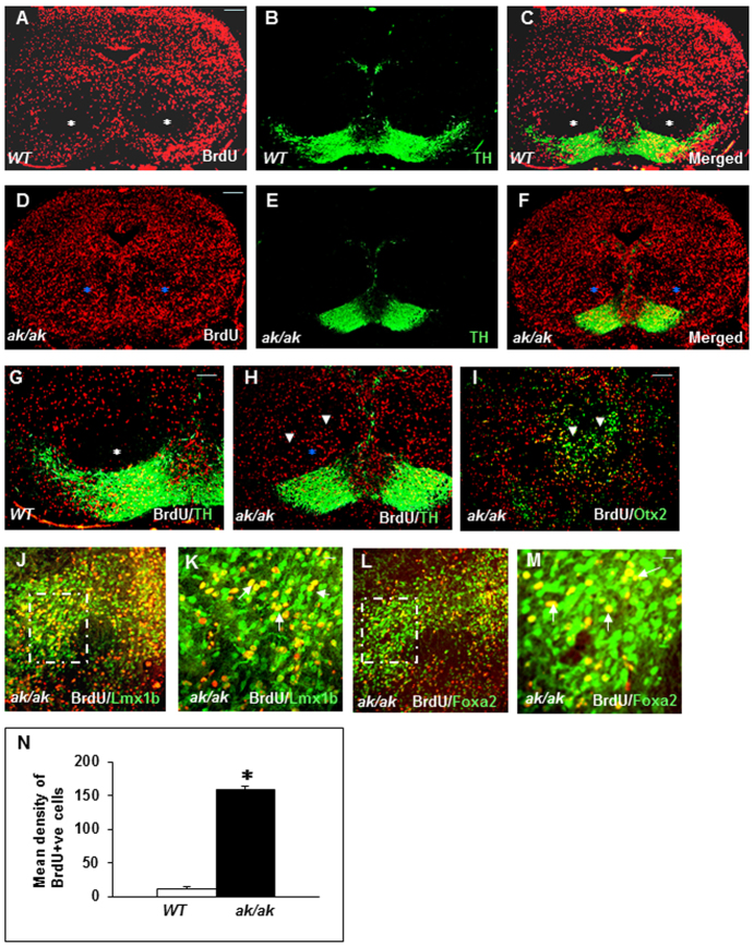
Abnormal neuronal migration and distribution of neurons in ak/ak mesencephalon. (A-M) The migrational profile of E11-labeled neuronal progenitors was analyzed in WT (A-C,G) and ak/ak mutant (D-F,H-M) mice at E17. White asterisks indicate areas free of BrdU-labeled cells in WT mesencephalon (A-C,G) and blue asterisks indicate abnormal cell clusters in the red nucleus of the ak/ak mutant (D-F,H). (G,H) Higher magnifications of C and F displaying failed perpendicular migration (arrowheads) in the ak/ak mutant. (I-M) Stalled cells in the ak/ak mutant are double positive for BrdU/Otx2 (arrowheads, I), BrdU/Lmx1b (J,K) and BrdU/Foxa2 (L,M). The boxed regions in J and L are magnified in K and M, respectively, and white arrows indicate double-positive cells. (N) Quantification of E11 BrdU-labeled cells distributed in the red nucleus (both hemispheres) of WT and ak/ak mutant (mean density of BrdU+ cells ± s.d.). A significant increase in BrdU+ cells was observed in the ak/ak mutant. *P<0.0001; n=5. Scale bars: 100 μm in A-J,L; 50 μm in K,M.

