
Fig. 1.
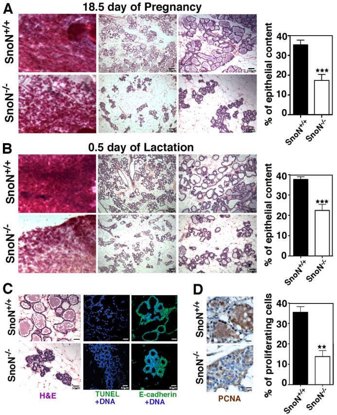
SnoN affects mammary alveolar development in mouse. (A,B) Alveolar atrophy is observed in the SnoN–/– mammary glands at day 18.5 of pregnancy (A) and day 0.5 of lactation (B). Left: whole-mount images from SnoN+/+ and SnoN–/– glands. Middle and right: Hematoxylin and Eosin (H&E) staining of mammary tissue sections at low (middle) and high (right) magnifications. The graphs to the right show the quantification of the epithelial content in three pairs of mice for day 18.5 pregnancy (P<0.0001) and in seven SnoN+/+ and six SnoN–/– mice at day 0.5 of lactation (P=0.0004). (C) H&E, TUNEL and E-cadherin staining at day 18.5 of pregnancy. (D) Pcna staining on alveolar structures at day 18.5 of pregnancy. Quantification of Pcna-positive nuclei in three pairs of glands (P=0.0140) is shown to the right. At least 1000 nuclei from each gland were counted. Error bars indicate the mean ± s.e.m. of three pairs of glands from each genotype.