
Fig. 5.
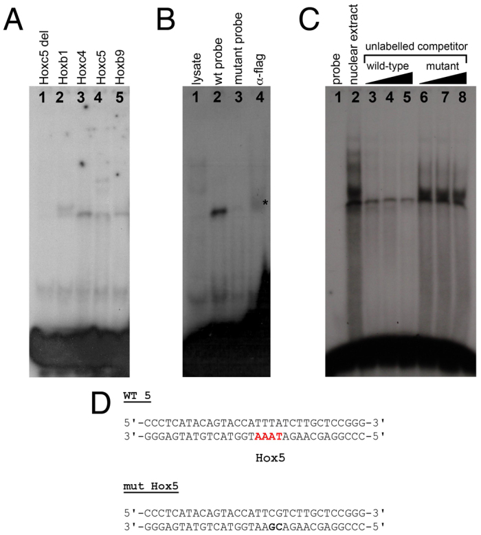
Direct binding of Hox proteins to the in-silico-predicted Hox binding sites. (A) Binding of in vitro translated mouse Hox proteins to the predicted Hox binding site 5 (Hbs5). Hoxc5 delHD provides a control. (B) Binding assay to mutant oligonucleotides and supershift assays for the predicted Hbs5. Lane 1, lysate control; lane 2, flag-tagged mouse Hoxc4 binding to labelled wild-type double-stranded oligonucleotide; lane 3, flag-tagged Hoxc4 fails to bind to labelled mutant double-stranded oligonucleotide; lane 4, supershift of the Hoxc4-Hbs5 complex with anti-flag antibody (asterisk). (C) Nuclear extracts obtained from prospective E9 forelimb regions were subjected to EMSA with labelled double-stranded oligonucleotide containing Hbs5. To test for the specificity of the shifted bands we performed competition analyses with excess unlabelled wild-type or mutant double-stranded oligonucleotide (100× molar excess, lanes 3, 6; 200×, lanes 4, 7; 500×, lanes 5, 8). (D) Double-stranded oligonucleotides used for EMSA contained mutated (mut Hox5, bold text) or wild-type (WT 5) Hbs5 (red).