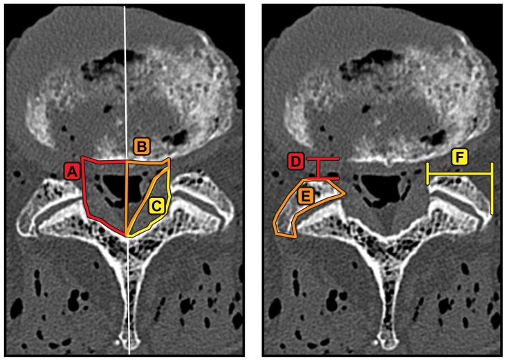
Figure 6.
Example of measurements made from reconstructed axial slices through the center of the intervertebral disc space aligned with the inferior endplate at the level of interest. (A) Bony canal area, (B) soft tissue canal area, (C) ligamentum flavum area, (D) lateral recess diameter, (E) facet area, and (F) facet width.
Notes: The spinal canal was defined to have a width equal to one-third of the left to right width of the intervertebral disc to avoid large variations in measurements. Left and right canal measurements were taken from the midline.

