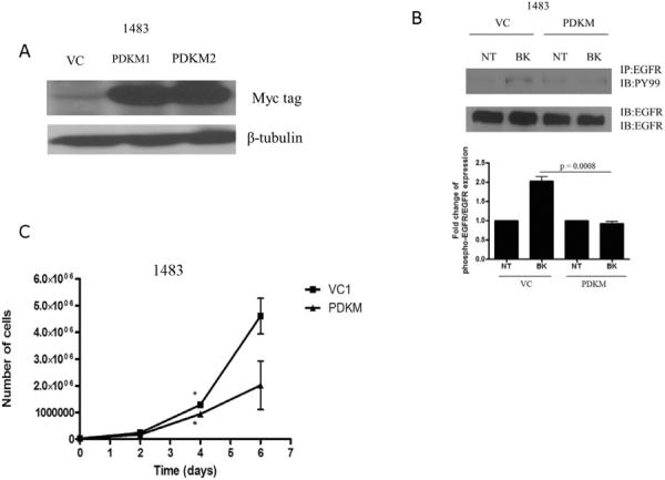
Figure 2. Kinase-dead PDK1 abrogates GPCR-mediated signaling and HNSCC growth.

(A) 1483 cells were stably transfected with pcDNA3.1 (vector control; VC) or pcDNA3-PDK1M (K110Q-kinase dead) and selected with neomycin for 2 weeks. Lysates were resolved by SDS-PAGE and probed for myc-tag and β-tubulin. (B) 1483 vector-transfected control (VC) and 1483 kinase-dead PDK1 (PDKM) cells were seeded, serum-starved for 72 hours and stimulated with BK (10 nM) for 5 minutes. Lysates were collected and immunoprecipitated with anti-phosphotyrosine antibody (P-EGFR) and immunoblotted for EGFR. Lysates were separately immunoblotted for EGFR to determine equal EGFR expression levels. Experiment was performed twice with similar results (C) 1483 VC and PDKM cells were seeded and assessed for growth by trypan blue dye exclusion on days 1, 3 and 5. Cell counts were graphed using GraphPad Prism Software (Day 5, p=0.039).
