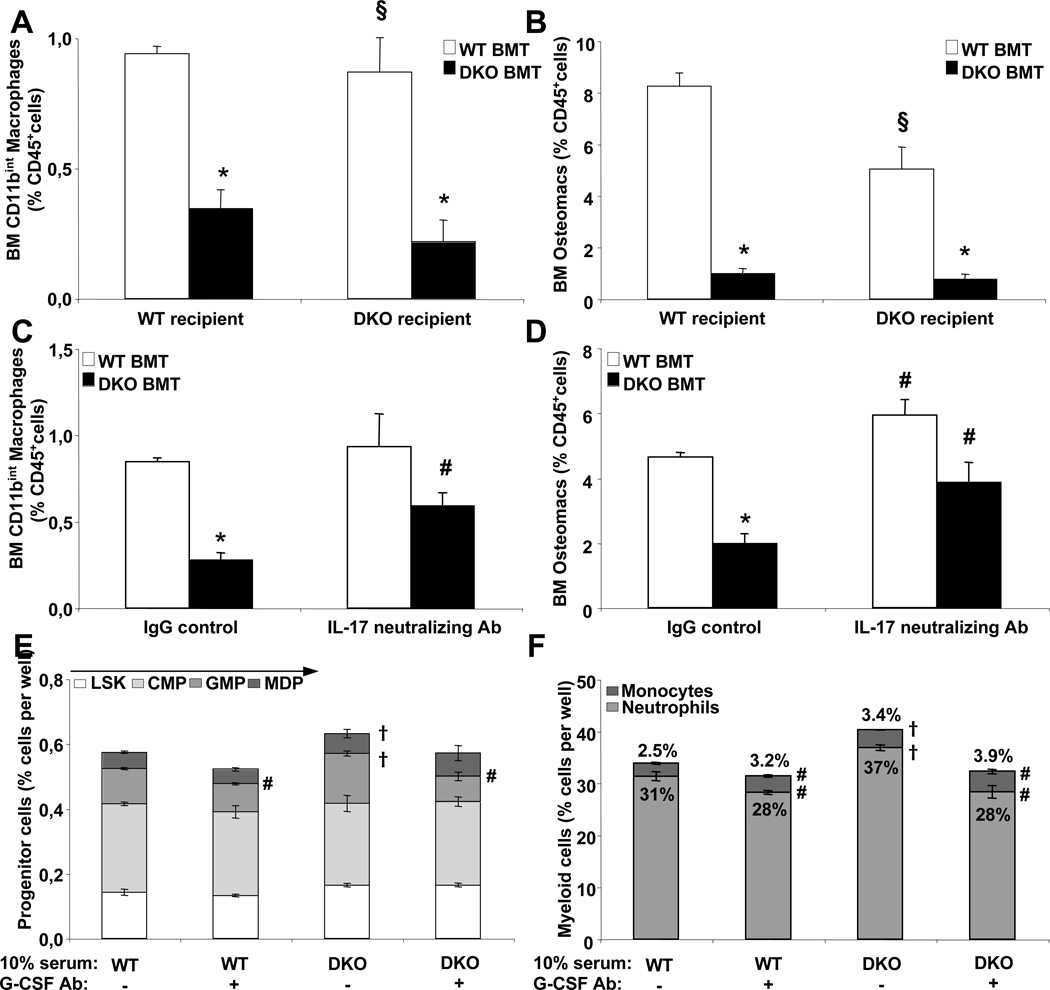
Figure 6. Depletion of BM macrophages in Abca1−/−Abcg1−/− mice.
Quantification of BM macrophage subsets by flow cytometry in WT and Abca1−/−Abcg1−/− recipient mice transplanted with either WT or Abca1−/−Abcg1−/− BM 8 weeks post-reconstitution (A and B) and in WT recipients transplanted with WT and Abca1−/−Abcg1−/− BM 14 weeks post-reconstitution and i.p injected with IgG control or 250µg IL-17 neutralizing antibody for 16h (C and D). Results are ± SEM of 5 to 6 animals per group. *P<0.05 vs. WT. §P<0.05 vs. Abca1−/−Abcg1−/− recipients transplanted with Abca1−/−Abcg1−/− BM. Quantification of hematopoietic progenitors (E), monocytes and neutrophils (F) in WT BM cultures grown for 48h in liquid culture in presence of 10% of the indicated serum and 50ng/mL G-CSF neutralizing antibody (+) or control non-specific IgG (−). Results are ± SEM of 3 independent experiments. #P<0.05 vs. IgG control. †P<0.05 vs. 10% WT serum.

