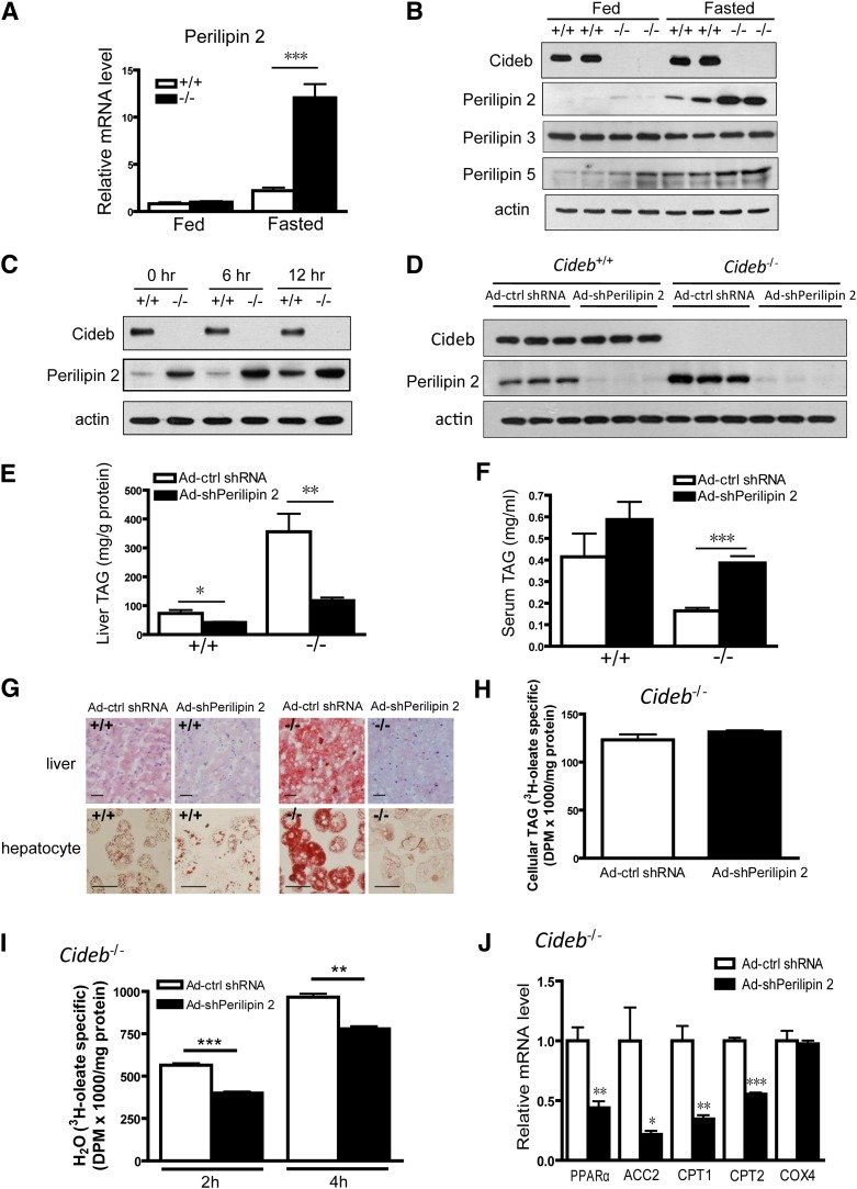
Fig. 3.
Knockdown of perilipin 2 in the livers of Cideb−/− mice results in decreased hepatic TAG accumulation. (A) Relative mRNA levels of perilipin 2 in the livers of 3-month-old wild-type (+/+) and Cideb−/− (–/–) mice (n = 5 each) under fed or fasting conditions. ***P < 0.001. Data are the means ± SEM. (B) Western blot showing increased perilipin 2 and perilipin 5 protein levels in the livers of Cideb−/− (–/–) mice under fasting conditions and no change in perilipin 3 levels in the absence of Cideb under either condition. The samples depicted are representative of four samples for each group. (C) Western blot showing increased perilipin 2 protein levels in Cideb−/− (–/–) hepatocytes in the presence or absence of 400 μm OA for the indicated duration. The samples depicted are representative of four independent experiments. (D) Perilipin 2 was knocked down in the livers of wild-type and Cideb−/− mice. Perilipin 2 shRNA was packaged into adenovirus (Ad-shPerilipin 2), and scrambled shRNA adenovirus was used as a negative control (Ad-ctrl shRNA). The viruses were introduced into 3-month-old Cideb−/− mice (n = 5 each) by tail-vein injection. The mice were sacrificed, and the livers were collected one week after injection. The samples depicted are representative of five samples for each group. (E) Knockdown of perilipin 2 decreased hepatic TAG levels in both wild-type (+/+) and Cideb−/− (–/–) mice (n = 5 each). *P < 0.05, **P < 0.01. Data are the means ± SEM. (F) Knockdown of perilipin 2 increased the plasma TAG levels in Cideb−/− (–/–) mice (n = 5 each). The serum was collected 12 h after fasting. ***P < 0.001. Data are the means ± SEM. (G) Representative photomicrographs of liver sections (upper panel) and isolated hepatocytes (lower panel) from Cideb−/− (–/–) and wild-type (+/+) mice stained with Oil Red O after treatment with Ad-shPerilipin 2 and control virus (Ad-ctrl shRNA). Scale bars, 50 μm. (H) Rates of TAG synthesis in Cideb−/− hepatocytes. Hepatocytes with perilipin 2 knockdown and control cells (n = 3 each) were incubated with radiolabeled [3H]oleic acid for 2 h. The TAG in the hepatocytes was extracted and evaluated by TLC. Data are the means ± SEM. (I) Decreased fatty acid β-oxidation rate in Cideb−/− hepatocytes with perilipin 2 knockdown compared with control cells. The hepatocytes (n = 3 each) were incubated with radiolabeled [3H]oleic acid for 2 h or 4 h. The 3H2O released into medium was extracted and evaluated. Data are the means ± SEM. **P < 0.01, ***P < 0.001. (J) Relative mRNA levels of PPARα, ACC2, CPT1, CPT2, and COX4 in the livers of Cideb−/− mice treated with Ad-shPerilipin 2 or Ad-ctrl shRNA (n = 5 each). The mice were fasted for 12 h. *P < 0.05, **P < 0.01, ***P < 0.001.

