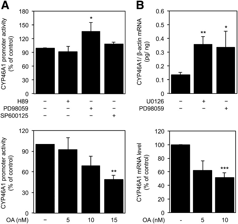
Fig. 8.
Effect of kinase/phosphatase chemical inhibitors on CYP46A1 basal expression. A: The 0.12pGL2 plasmid was transfected into SH-SY5Y cells. Twenty-four hours after transfection, cells were incubated with 5 μM H89, 10 μM PD98059, 10 μM SP600125, or with the indicated concentrations of OA for 16 h. Normalized luciferase activities were expressed as mean values ± SEM of duplicates for a minimum of three independent experiments. B: Real-time PCR analysis of CYP46A1 steady-state mRNA transcripts level in SH-SY5Y and NT2N cells treated, respectively, with 10 μM U0126 or 10 μM PD98059 and with the indicated doses of OA for 16 h. Values were normalized to the internal standard β-actin. Data represent means ± SEM of at least three independent experiments and are expressed as percentage of induction relative to vehicle-treated cells (NT2N) or as pg of CYP46A1 per ng of β-actin (SH-SY5Y) (*P < 0.05; **P < 0.01; ***P < 0.001).

