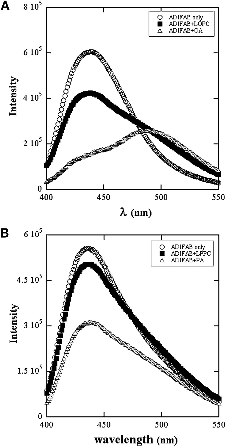
Fig. 1.
Fluorescence emission spectra of ADIFAB excited at 386 nm in (A) Hepes buffer without (○) and with 1 μM each of LOPC(■) and OA(△). B: Hepes buffer without (○) and with 1 μM each of LPPC(■) and PA(△). Acrylodan is displaced and its fluorescence emission shifts to the red region of the spectrum upon binding of LPC and FA to the ADIFAB protein.

