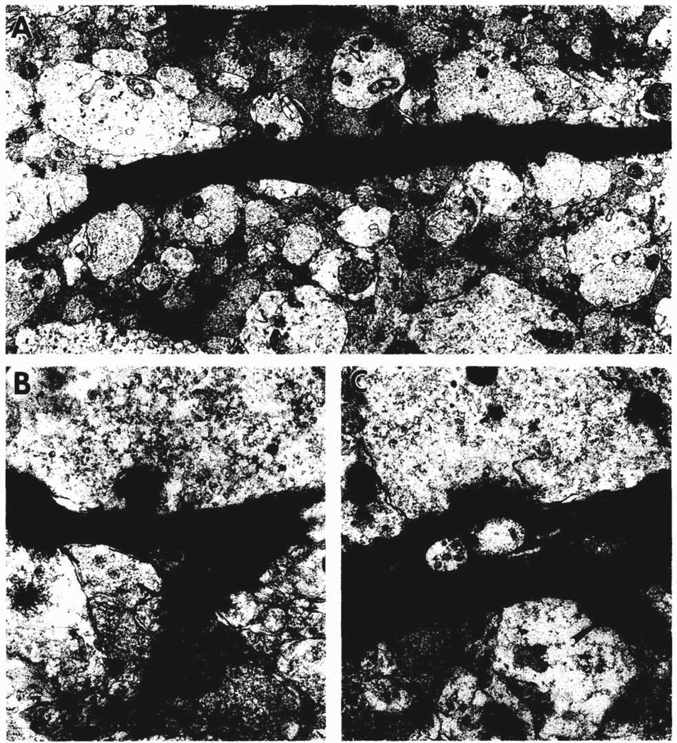
Fig. 3.
Electron micrographs of dendrites in stratum lucidum of CA3b that arise from degenerating neurons in the pyramidal cell layer. A shows two longitudinally-sectioned, degenerating dendrites (d and arrow) found in the neuropil that also contains many normal cross-sectioned dendrites and large axon terminals. B shows a stubby spine from the dendrite at the top of A. Two axon terminals with round synaptic vesicles form synapses (probably asymmetric, arrows) with the electron dense dendrite (left) and a stubby spine (right). C shows a pedunculate spine (large arrow) and an axodendritic synapse (small arrow) formed by a large axon terminal with round synaptic vesicles and a few dense core vesicles, similar in appearance to a developing mossy fiber axon terminal. A = ×10,000; B = ×37,000; C = ×28,000.

