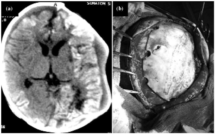
Fig. 2.

Patient under early DC procedure. (a) Satisfactory evolution. MRI shows the skull defect and post-traumatic parenchyma changes. (b) Third phase of reconstruction and definitive close with the patient's osseous graft from bone bank. (Photo: Author).
