
Fig. (4). Modification to the 2’-backbone and 3’-termini in saRNA.
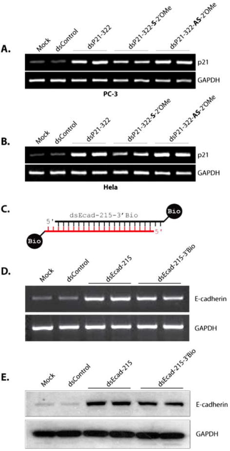
(A-B) PC-3 (A) and HeLa (B) cells were transfected with 50 nM concentrations of dsControl, dsP21-322, dsP21-322-S-2’OMe, or dsP21-322-AS-2’OMe for 72 hours. Mock samples were transfected in the absence of saRNA. Expression of p21 and GAPDH were evaluated by standard RT-PCR. (C) Schematic representation of dsEcad-215-3’Bio possessing biotin covalently linked to both 3’-termini. The antisense strand is in red, while the sense strand is black. (D) PC-3 cells were transfected at 50 nM dsControl, dsEcad-215, or dsEcad-215-3’Bio for 72 hours. Expression of E-cadherin and GAPDH mRNA levels were evaluated by standard RT-PCR. (E) Induction of E-cadherin protein was confirmed by immunoblot analysis. GAPDH was also detected and served as a loading control.