Figure 6.
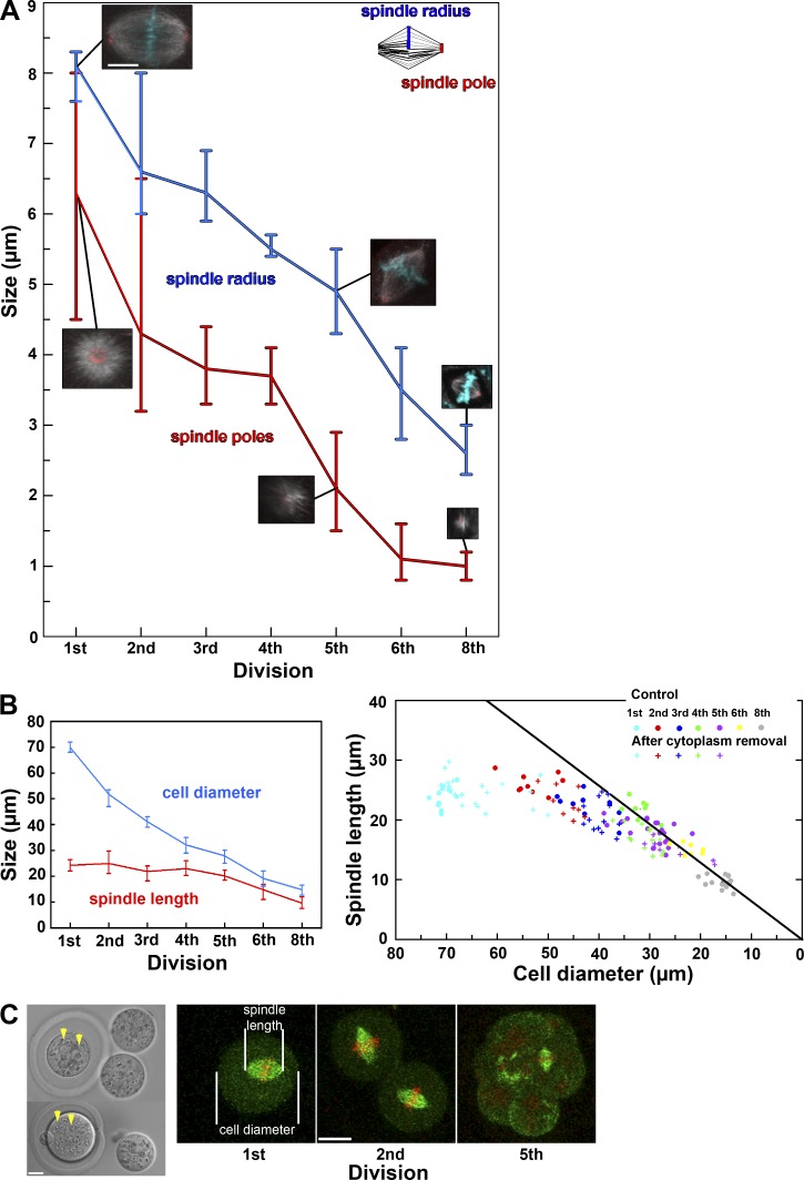
Gradual change in spindle characteristics and establishment of the spindle length regulation during mouse preimplantation development. (A) Progressive change in radius and diameter of the spindle in the embryos at consecutive developmental stages. Insets show a representative image of the spindle and its pole at each stage. Bar, 10 µm. (B, left) Change in the mean size of the spindle and of the cell in the embryos at consecutive developmental stages. (right) Cell diameter and spindle length are plotted as colored circles for individual embryos at different developmental stages, illustrating that their ratio (slope of the black line) remains constant from the fourth to eighth division. Those for experimentally micromanipulated embryos are shown as colored crosses. (C, left) Experimentally micromanipulated zygotes in which two thirds (top) or half (bottom) of the cytoplasm was removed. Note that two pronuclei are visible (yellow arrowheads) after cytoplasm removal. Metaphase spindle and measurement of its size and cell size by live imaging of the micromanipulated embryos during the subsequent divisions. All pictures are projected images of 4.5-µm stacks. Bars, 20 µm. In A and B, the vertical bars indicate the range of values.

