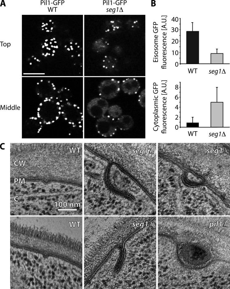
Figure 1.
Seg1 is required for proper eisosome architecture. (A) Confocal images of Pil1-GFP in wild-type (WT) and seg1Δ cells. Representative top views and mid sections are shown. Bar, 5 µm. (B) Quantification of Pil1-GFP signal per eisosome (eisosome GFP fluorescence) and Pil1-GFP signal in the cytoplasm (cytoplasmic GFP fluorescence) in WT and seg1Δ cells. A.U., arbitrary units. Error bars indicate standard deviations. (C) Electron micrographs of WT, seg1Δ, and pil1Δ cells. CW, cell wall; PM, plasma membrane; C, cytoplasm.

