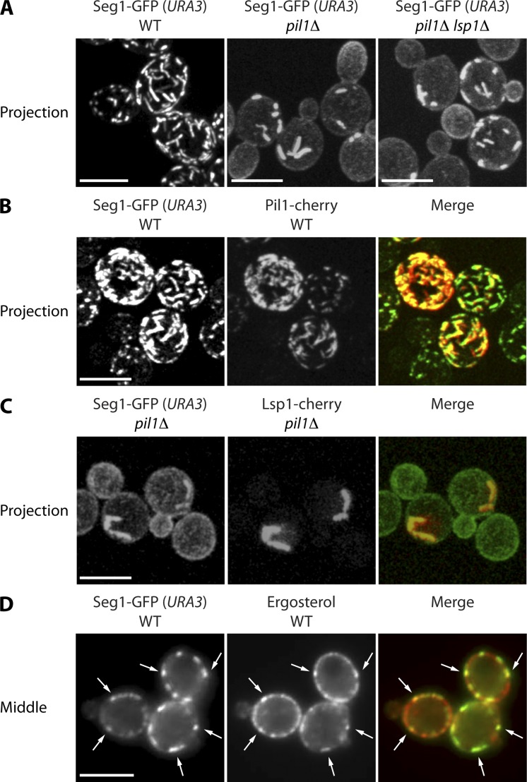
Figure 6.
Seg1 rods form without Pil1 and contain eisosome components. (A) Projections from confocal stacks of wild-type (WT), pil1Δ, and pil1Δ lsp1Δ cells lacking endogenous Seg1 and expressing Seg1-GFP from the URA3 locus. (B) Projections of WT cells expressing Pil1-cherry, lacking endogenous Seg1, and expressing Seg1-GFP from the URA3 locus. (C) Projections of pil1Δ cells expressing Lsp1-cherry, lacking endogenous Seg1, and expressing Seg1-GFP from the URA3 locus. (D) Epifluorescence images of WT cells lacking endogenous Seg1, expressing Seg1-GFP from the URA3 locus, and stained with filipin to visualize ergosterol. Arrows indicate colocalization of Seg1-GFP and filipin. Bars, 5 µm.

