Figure 1.
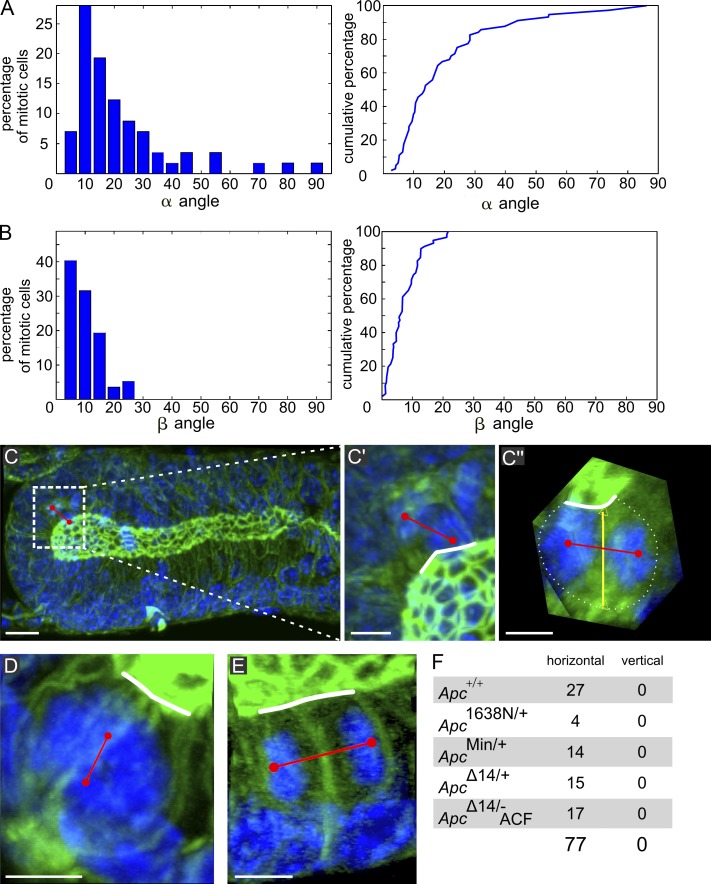
Spindle orientation in the tubular part and the semispherical parts of colon crypts. (A and B, left) α and β angles in 67 mitotic cells in wt crypts of six animals. (A and B, right) Cumulated percentages: 80% of α angles are ≤30°, but ∼20% display angles up to 90°. Nearly 100% of β angles are ≤20°. (C) MIP of optical sections of a crypt comprising a telophase. (C′ and C′′) Zoomed-in images of the region of interest defined by the dotted rectangle in C, where C′′ is 3D rotated to display the spindle axis perpendicular to the observer. Red line shows spindle axis; white line shows apical surface; yellow line links the center of apical surface and basal pole. In C′, the spindle appears nearly perpendicular to the apical surface, whereas C′′ shows it is in fact parallel (complete 3D examination shown in Video 2). (D) MIP of a prometaphase displaying a spindle axis perpendicular to the apical surface (3D viewing shown in Video 3). (E) MIP of a telophase in an ApcMin/+ mouse with the spindle parallel to the apical surface. (F) Number of meta/telophases at the crypt base exhibiting horizontal versus vertical spindle orientation in wt (Apc+/+), Apc1638N/+, ApcMin/+, and ApcΔ14/+ crypts and in ACF of ApcΔ14/− mice (ApcΔ14/− ACF). Bars: (C) 10 µm; (C′–E) 5 µm.

