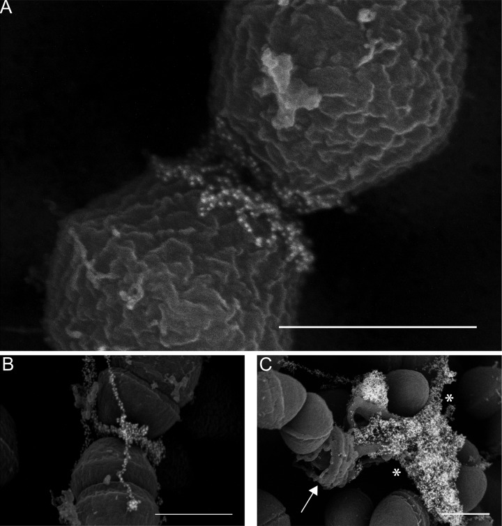
FIG 3 .
(A and B) Immuno-SEM micrographs demonstrating localization of the eDNA probe near the E. faecalis septum. (C) Endogenous lysis of cells in an older (48-h) biofilm display an entirely different morphology from that seen in early biofilms, as DNA (asterisks) is released from a ruptured cell (arrow). Bars, 500 nm.

