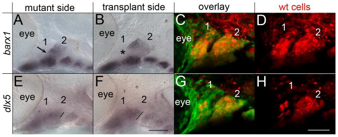
Fig. 6.
Neural crest cells require the reception of Hh signaling for proper dorsal/ventral patterning. (AH) 36 hpf fli1:EGFP;smo-/- embryos imaged following the transplantation of neural crest cells from fli1:EGFP;smo+/+ donors. The fli1:EGFP;smo+/+ donors were injected with Alexa 568 dextran to visualize the transplanted cells (in red). The mutant side, not receiving the transplant, shows dorsal/ventral patterning defects, with the fusion of the dorsal and ventral domains of barx1 (A, arrow) and a reduced extent of dlx5a expression (E, line). (B-D) Neural crest cells wild type for smo restore the intermediate barx1 free expression domain (B, arrowhead). (F-H) Transplanted neural crest cells also increase the extent of dlx5a expression (E, line). (C & G) Show the overlay of fli1:EGFP and Alexa 568 while (G & H) are the same embryos showing just the transplanted cells. Note that in this embryo there is poor contribution of neural crest cells to the first pharyngeal arch, although the second arch is highly populated with transplanted cells. The first two pharyngeal arches are labeled. Anterior is to the left; dorsal is up in all images. Scale bar=50μm.

