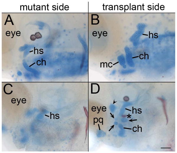
Fig. 9.
Reception of Hh signaling by the endoderm can reorganize the facial skeleton. (A & C) Mutant and (B & D) transplant side of smo mutant embryos that have received wild-type endoderm transplants and were stained with Alcian Blue/Alizarin Red at 4 dpf. (A & B) In embryos that produce cartilage, these transplants result in differently shaped cartilage elements. (C & D) In the most extensive reorganization of the pharyngeal skeleton, (C) the mutant side of the embryo has a single cartilage nodule in the appropriate location for the hyosymplectic (hs). (D) The transplanted side of the same embryo has a nodule in the correct location for the hyosymplectic with an attached opercle bone (asterisk). A cartilage nodule is also in the appropriate location for the ceratohyal cartilage (ch) and there are three nodules (arrows) in the location for the interhyal or symplectic rod. Adjacent to the eye there is a nodule in the appropriate location for the palatoquadrate (pq) and an ectopic nodule is located just posterior to the eye (arrowhead). Anterior is to the left in all images. Scale bar=50μm.

