
Figure 1. Preparation and positioning of a transverse section.
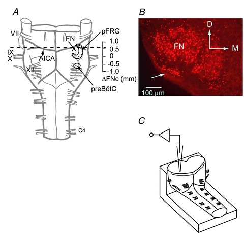
A, ventral view of a brainstem–spinal cord preparation from a neonatal rat. The preparation was cut at the level of the dotted line. B, rostral cut surface view of Phox2b-immunoreactive (-ir) cells at a level 0.5 mm rostral to the caudal end of the facial nucleus (ΔFNc). Phox2b-ir cells (red) are located in the ventral parafacial region (arrow) and in the facial nucleus. C, schematic representation of the preparation in the recording chamber. The preparation was fixed onto a rubber block by pins with the cut surface facing up for whole-cell recordings of the parafacial region via the cut surface of the cross section. AICA, the anterior inferior cerebellar artery; FN, facial nucleus; pFRG, parafacial respiratory group; preBötC, preBötzinger complex; VII–XII, cranial nerves; C4, the fourth cervical ventral root; D, dorsal; M, medial.