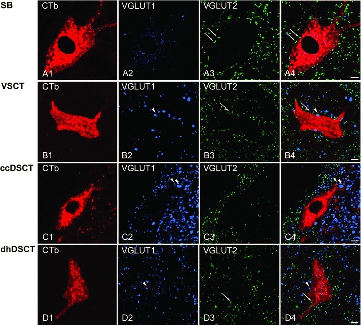
Figure 9. Immunohistochemical characteristics of VGLUT1 and VGLUT2 axon terminals in contact with retrogradely labelled spinocerebellar neurons.

A1–A4, B1–B4, C1–C4 and D1–D4, single optical sections through the cell bodies of representative SB, VSCT, ccDSCT and dhDSCT neurons illustrating the contacts made by VGLUT1 (blue) and VGLUT2 (green) immunoreactive boutons indicated by arrowheads and arrows, respectively. The cell body and dendrites of retrogradely labelled cells are in red. Note differences in the density of VGLUT1- and VGLUT2-immunoreactive terminals within different regions of the grey matter shown in A–D and especially the high density of VGLUT1 and low density of VGLUT2 within Clarke's column shown in C. Scale bar in A–D, 10 μm.
